简体中文
简体中文
 English


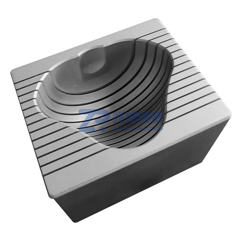
Made of high-quality stainless steel series, it can be rapidly heated, rapidly cooled, autoclaved, and sterilized by alcohol immersion. It is durable, and the quality of repeatedly sliced sections can be well guaranteed.
Two common slicing forms are used for brain slicing molds:
Longitudinal sections parallel to the midline of the brain (sagittal) are called coronal sections; transverse sections perpendicular to the midline of the brain (coronal) are called sagittal sections. The slice thickness is 1mm or 0.5mm. For larger animals (monkeys, beagles, rabbits, etc.), a slice thickness of 2mm is commonly used.
Ordering Information
| Model | Name | Body Weight (kg) | Slicing Direction | Slice Thickness (mm) |
| Z5185 | Cynomolgus Monkey/Rhesus Monkey Cerebellar Brain Mold | 4-11 | Coronal | 2.0 |
| Z5186 | Cynomolgus Monkey/Rhesus Monkey Cerebellar Brain Mold | 3-7 | Sagittal | 2.0 |
Usage Reference
Compatible with: 235-Triphenyltetrazolium Chloride (TTC) Staining Solution
1. Take the brain tissue sample to be tested (generally, the brain can be taken directly after anesthesia or after perfusion with normal saline), and after taking it: quick-freeze at -20℃ for 20~30min to facilitate slicing.
2. Slice the brain tissue to be tested; generally, the thickness of animal samples is 0.5~2mm.
3. Put the slices into TTCStain (2%) and incubate in the dark for 25~35min.
4. Fix the slices in 4% paraformaldehyde or 10% neutral formalin for 4~24h.
5. Blot dry the water on the tissue surface, and use image analysis systems such as IPP to measure the cerebral infarction area and calculate the cerebral infarction volume.